Media Summary: CellSens 3D deconvolution Fluorecence Imaging This video was recorded by the Live Cell Imaging facility at the Karolinska Institute in Sweden during the LCI course 2026. ImageJ macro for iterative 2D deconvolution for 3D image stack
3d Deconvolution Fluorescent Z Stacking - Detailed Analysis & Overview
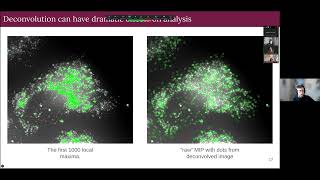
CellSens 3D deconvolution Fluorecence Imaging This video was recorded by the Live Cell Imaging facility at the Karolinska Institute in Sweden during the LCI course 2026. ImageJ macro for iterative 2D deconvolution for 3D image stack The Olympus cellSens platform creates a uniquely personal and intuitive imaging experience based on the operator's preferred ... - Describes the basics of Media Cybernetics' AutoQuant image Mouse fibroblast spheroid (NIH 3T3, 500 cells) was imaged in a INCell Analyzer 2500HS ( ). Magnification:4X.


![FIJI (ImageJ): 3D Image Deconvolution [Theoretical PSF]](https://i.ytimg.com/vi/6AvMjiVMRPA/mqdefault.jpg)